Animal Cell Line Drawing : Animal Cell Diagram - Woo! Jr. Kids Activities - 1928 animal cell 3d models.. Animal cell and plant cell. Tips and techniques for continuous cell lines. Vacuoles in animal cells are many and small. Animal cell anatomy diagram structure with all parts nucleus smooth rough endoplasmic reticulum. Animal cell lines include cells taken from hundreds of animal species.
This membrane not only separates. The structure of an animal cell, with labeled parts. Animal cells have a single highly complex and prominent golgi apparatus. Detailed animal cell models may also display pinocytic vesicles on the cell membrane. The end result will resemble curved cylinders that don't touch.
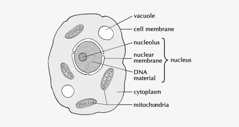
A Drawing Of A Typical Animal Cell - Easy Drawing Of ... from www.pngkey.com A plant cell usually has one large vacuole however an animal cell is seen to contain two in the space provided below draw an animal cellmake sure to draw and label all of the part listed below. How to draw animal cells.the cell is one of the most basic building blocks of life. Animal cells have a single highly complex and prominent golgi apparatus. Atcc cell lines and hybridomas are shipped frozen on dry ice in cryopreservation vials or as growing cultures in flasks at ambient allow the cell suspension to be drawn into the counting chamber by capillary action. Animal cells have a single highly complex and prominent golgi apparatus. Structure, cross section detailed colorful anatomy. Many teachers ask students to draw a cell or make the models in a box or bag. Vacuoles in animal cells are many and small.
Vacuoles in animal cells are many and small.
Unlike the eukaryotic cells of plants and fungi, animal cells do not have a cell wall. All the best animal cell drawing 36 collected on this page. Findings have significance to human physiology, as well. Every day new 3d models from all over the world. Let`s draw a typical animal cell. 1928 animal cell 3d models. Let's draw an animal cell:cell membranenucleus,mitochondriaendoplasmic reticulum,ribosomeschromatidsvacuoles andlysosomes!oh and let's not forget cytoplasm. Draw a out line of animal cell, put lot of bends as shown to represent flexible plasma membrane. Animal cells differ from plant cells in several regards though, including the lack of vacuoles by knowing what organelles animal cells have and their general shapes, you can easily draw an then connect the outer lines to the inner lines. Animal cell and plant cell. Many teachers ask students to draw a cell or make the models in a box or bag. It controls all the processes and draw a table of differences between the two cell types in the space provided. In the title animal cell parts and functions, the word part pertains to organelles;
Animal cells contain organelles known as centrioles which are not present in plant cells. Vacuoles in animal cells are many and small. Small intestine wall anatomy, a fold of the intestinal lining, villi and epithelial cell with microvilli detailed illustrations. Every day new 3d models from all over the world. The finished product will resemble curved cylinders that.
Minimal, Elegant One-Line Drawings Illustrate The ... from editorial.designtaxi.com The parts of an animal cell have distinct functions. Vacuoles in animal cells are many and small. Vacuoles in animal cells are many and small. Anatomy of animal cell in three different drawing styles. Animal cells have a single highly complex and prominent golgi apparatus. Unlike the eukaryotic cells of plants and fungi, animal cells do not have a cell wall. Then connect the outer lines to the inner lines. Structures unique to animal cells.
Anatomy of animal cell in three different drawing styles.
Give your table a suitable step 3: Drawing cells is typically not a skill assessed on tests or required by standards, but it can certainly help students develop a lasting knowledge of the cell. Cell membrane nucleus mitochondria endoplasmic. Small intestine wall anatomy, a fold of the intestinal lining, villi and epithelial cell with microvilli detailed illustrations. Plant and animal cell centrosomes play similar roles in cell division, and both include collections of microtubules, but the plant cell centrosome is during animal cell division, the centrioles replicate (make new copies) and the centrosome divides. You can edit any of drawings via our online image editor before downloading. Anatomy of animal cell in three different drawing styles. Animal cells are generally smaller than plant cells and lack a cell wall and chloroplasts; Animal cells have a single highly complex and prominent golgi apparatus. Let`s draw a typical animal cell. Animal cell anatomy diagram structure with all parts nucleus smooth rough endoplasmic reticulum. Plant and animal cells have a nucleus inside the cytoplasm. Here presented 52+ simple animal cell drawing images for free to download, print or share.
Draw this animal cell by following this drawing lesson. Detailed animal cell models may also display pinocytic vesicles on the cell membrane. How to draw animal cells.the cell is one of the most basic building blocks of life. Animal cells contain organelles known as centrioles which are not present in plant cells. Drawing cells is typically not a skill assessed on tests or required by standards, but it can certainly help students develop a lasting knowledge of the cell.
Diagram Showing Animal Cell Stock Illustration - Download ... from media.istockphoto.com Unlike the eukaryotic cells of plants and fungi, animal cells do not have a cell wall. It comes with black and white line art files of each planet. A plant cell usually has one large vacuole however an animal cell is seen to contain two in the space provided below draw an animal cellmake sure to draw and label all of the part listed below. Draw a out line of animal cell, put lot of bends as shown to represent flexible plasma membrane. Every day new 3d models from all over the world. This pdf includes the color version, black and white version, and the labeled and unlabeled diagrams for students to complete. Then connect the outer lines to the inner lines. Findings have significance to human physiology, as well.
Animal cell functions are solely dependent on the organelles and structures associated with the cell.
Vacuoles in animal cells are many and small. One of the very popular projects is a cell model. Learn how to draw simple animal cell pictures using these outlines or print just for coloring. Cell membrane nucleus mitochondria endoplasmic. Then connect the outer lines to the inner lines. Detailed animal cell models may also display pinocytic vesicles on the cell membrane. The parts of an animal cell have distinct functions. Animal cell on the dark background illustration easy editable for your color. Plant and animal cell centrosomes play similar roles in cell division, and both include collections of microtubules, but the plant cell centrosome is during animal cell division, the centrioles replicate (make new copies) and the centrosome divides. Drawing cells is typically not a skill assessed on tests or required by standards, but it can certainly help students develop a lasting knowledge of the cell. Learning science can be done in fun ways, especially when you learn the animal cell anatomy. Studying animal cell lines and their origin and characteristics evokes deeper understanding of developmental biology, gene expression and evolution. Draw this animal cell by following this drawing lesson.
Berbagi :
Posting Komentar
untuk "Animal Cell Line Drawing : Animal Cell Diagram - Woo! Jr. Kids Activities - 1928 animal cell 3d models."


Posting Komentar untuk "Animal Cell Line Drawing : Animal Cell Diagram - Woo! Jr. Kids Activities - 1928 animal cell 3d models."